
Introduction
Gallbladder polyps are usually incidental findings diagnosed during abdominal ultrasound exams or during cholecystectomy. They usually do not present symptoms, but occasionally they can cause discomforts similar to those caused by gallstones.
Most of these lesions are not neoplastic, but rather hyperplastic or represent lipid deposits.
With the widespread use of ultrasound, polypoid lesions in the gallbladder are being increasingly detected. However, often the image is not enough to rule out the possibility of neoplasia or pre-malignant adenomas. In this article, we will review the clinical importance and differential diagnosis of gallbladder polyps.
Classification
Polypoid lesions in the gallbladder can be categorized as benign or malignant. Benign lesions can be subdivided into neoplastic and non-neoplastic.
Non-neoplastic benign polyps
The most common benign non-neoplastic lesions are cholesterol polyps, followed by adenomyomatosis and inflammatory polyps.
- Cholesterol polyps and cholesterosis:
- it is a benign condition characterized by the accumulation of lipids in the mucosa of the gallbladder wall.
- they are the most common types of gallbladder polyps, reaching up to 10% or more.
- It can be of the diffuse or polypoid type.
- The term cholesterosis refers to the diffuse type, which is usually diagnosed incidentally during cholecystectomy, causing the appearance of a “strawberry gallbladder” due to the contrast it makes with the gallbladder mucosa.
- Cholesterol polyps are the polypoid form of cholesterosis, being the most common gallbladder polyp, usually diagnosed incidentally on ultrasound.
- Although usually asymptomatic, in some patients it can cause symptoms and complications similar to those caused by gallstones.
- Adenomyomatosis:
- it is an abnormality of the gallbladder characterized by excessive growth of the mucosa, thickening of the muscular wall and intramural diverticula.
- The prevalence of gallbladder adenomyosis is low, but it appears to have a higher prevalence in women than in men.
- Inflammatory polyps
- Inflammatory polyps are the least common non-neoplastic polyps.
- They appear as sessile or pedunculated and are composed of granulation and fibrous tissue with plasma cells and lymphocytes.
- The polyps are usually 5 to 10 mm in diameter, although inflammatory polyps larger than 1 cm have been described


Neoplastic benign polyps
- Adenomas:
- Adenomatous polyps of the gallbladder are the most common benign neoplastic lesions. Although the true incidence is unknown, in most series it is less than 0.5 percent.
- Gallbladder adenomas are benign epithelial tumors composed of cells that resemble the epithelium of the bile ducts.
- The risk of cancer increases with the size of the polyp, with larger adenomatous polyps having a risk of malignancy.
- Others — Other neoplastic lesions of the gallbladder such as fibromas, lipomas and leiomyomas, are rare. The natural history of these polyps is not well defined.
Malignant polyps:
- Most malignant polyps in the gallbladder are adenocarcinomas.
- The adenocarcinomas of the gallbladder are much more common than gallbladder adenomas, unlike the colon, where adenomas are much more common than adenocarcinomas.
- Squamous cell carcinoma, mucinous cystadenoma and gallbladder adenoacanthomas are rare
CANCER RISK
Most gallbladder polyps are benign, and most benign polyps, with the exception of adenomas, do not have malignant potential. The overall risk of gallbladder cancer in patients with gallbladder polyps appears to be low.
- In a large cohort study with over 35,000 adults with gallbladder polyps diagnosed by USG, 0.053% had gallbladder cancer, similar to the population without polyps (0.054%). [ref]
- The risk of progression to neoplasia varies according to the size of the polyps, occurring in 128/100,000 people for polyps > 10mm, but only in 1.3/100,000 people for polyps < 6mm.
Established risk factors for cancer
- Polyp size — The incidence of gallbladder cancer varies from 43 to 77% in polyps larger than 1 cm and 100% in polyps larger than 2 cm.
- Sessile polyp — sessile polyps are an independent risk factor for malignancy, with a 7x higher risk of gallbladder cancer. [ref]
- Age > 60 years: this is the cut-off adopted in guidelines for risk stratification and treatment guidance.
- Others: Indian ethnicity, primary sclerosing cholangitis
Conditions with uncertain risk
- Concomitant gallstones
- Adenomyomatosis — There is no evidence that the presence of adenomyosis increases the risk of gallbladder cancer. If the risk is increased, the magnitude of the increase appears to be small.
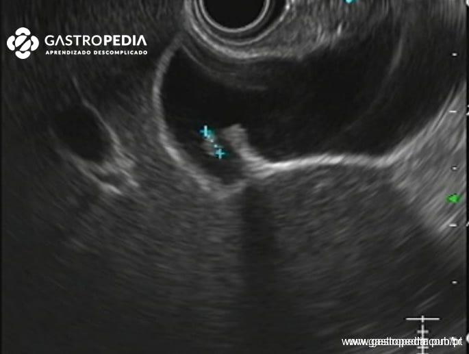
DIAGNOSIS
Gallbladder polyps are usually discovered incidentally on abdominal ultrasound exams. None of the available imaging modalities can unequivocally distinguish benign from malignant polyps. This can only be confirmed by histopathology after cholecystectomy.
Characteristics of gallbladder polyps on abdominal ultrasound:
- They can be single or multiple
- Sensitivity 84% and specificity 96% (meta-analysis with 16,260 patients)
- CHOLESTEROL POLYPS are usually multiple, homogeneous, polypoid and pedunculated, with echogenicity greater than the liver parenchyma.
- They may or may not contain hyperechoic points.
- Cholesterol polyps usually measure less than 1 cm.
- In contrast to cholesterol polyps, diffuse cholesterosis does not have specific ultrasonographic findings, and its diagnosis is usually made after surgery.
- ADENOMAS are homogeneous lesions, isoechoic in relation to the liver parenchyma, have a smooth surface and usually do not have a pedicle.
- The sessile morphology and focal thickening of the gallbladder wall greater than 4 mm are risk factors for malignancy.
- ADENOCARCINOMAS are homogeneous or heterogeneous polypoid structures that are usually isoechoic in relation to the liver parenchyma.
- The ADENOMYOMATOSIS can also cause a diffuse thickening with round anechoic f
Professor Livre-Docente pela Faculdade de Medicina da Universidade de São Paulo
Médico Endoscopista do Instituto do Câncer do Estado de São Paulo (ICESP)
Médico Endoscopista do Hospital Alemão Oswaldo Cruz
Emerging Star pela WEO
